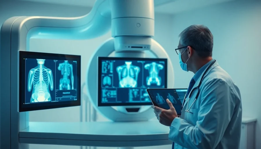
What is Digital X-Ray Equipment?
Introduction to Digital X-Ray Technology
Digital x-ray equipment is a transformative technology in the realm of diagnostic imaging, offering enhanced accuracy, efficiency, and ease of use compared to traditional analog x-ray systems. Unlike classic film-based radiography, digital x-ray captures images electronically, providing immediate access to high-quality images essential for accurate diagnosis and treatment decisions. This technology leverages various digital detectors such as flat-panel detectors or charge-coupled devices (CCDs) to capture and convert x-ray photons into digital signals, significantly improving the workflow in medical practices.
The evolution of digital x-ray equipment has revolutionized the radiographic landscape by making procedures quicker and more efficient. It not only enhances the quality of the images but also reduces the radiation dose to the patient, a critical factor in modern healthcare.
Benefits of Digital X-Ray Systems
The transition from traditional film x-rays to digital x-ray systems comes with a multitude of benefits that enhance diagnostic capabilities and improve patient outcomes. Key advantages include:
- Instant Image Retrieval: Digital x-ray systems eliminate the waiting time for film development, allowing healthcare providers to receive images almost instantaneously. This expediency can significantly speed up clinical decisions, particularly in emergency situations.
- High Image Quality: Digital x-rays provide superior image resolution and clarity. Advanced processing can enhance visibility of details that might be missed in film-based systems, leading to better diagnostic accuracy.
- Lower Radiation Dose: Digital x-ray technologies typically require lower doses of radiation for image capture, thus minimizing the exposure risk to patients while maintaining image quality.
- Flexible Storage and Transmission: Digital images can be easily stored, retrieved, and transmitted electronically, facilitating better record-keeping and enabling remote consultations among healthcare professionals.
- Environmental Benefits: Eliminating the need for film and chemicals associated with traditional development methods leads to a reduced environmental footprint, making digital x-ray a more sustainable option.
Key Components of Digital X-Ray Equipment
A digital x-ray system consists of several integral components working seamlessly together to produce high-quality diagnostic images. Understanding these components is crucial for selecting and maintaining the equipment effectively:
- X-Ray Tube: This component generates x-rays by using electricity to heat a filament, producing electrons which collide with a target material and generate x-ray photons.
- Image Receptor: The image receptor can be either a flat-panel detector or a CCD that captures the x-ray photons. This component converts the x-rays into electrical signals which are processed into images.
- Image Processing Software: Sophisticated software allows for image enhancement, manipulation, and analysis. This software includes features for adjusting contrasts, zooming in on specific areas, and identifying abnormalities.
- Workstation: The workstation is the hub where images are viewed, manipulated, and stored. It often includes features for networking with other systems, enabling easy access and sharing of images among healthcare providers.
- Radiation Safety Devices: Modern digital x-ray systems are equipped with safety devices to minimize radiation exposure for both patients and staff, emphasizing the importance of patient safety in the diagnostic process.
Understanding the Operation of Digital X-Ray Equipment
How Digital X-Rays Work
The operation of digital x-ray equipment is grounded in fundamental principles of physics and advanced technology. The process begins when the x-ray tube emits x-ray beams directed towards the patient, penetrating body tissues differently depending on their density.
The image receptor captures the exiting x-rays after they pass through the patient. For instance, denser areas like bones absorb more x-ray energy than softer tissue, resulting in varying shades of gray in the captured image. This data is transformed into a digital format for analysis through an analog-to-digital converter, allowing for precise imaging.
Common Techniques in Digital Radiography
Digital x-ray equipment supports various radiographic techniques that enhance diagnostic capabilities. Some common techniques include:
- Projection Radiography: This is the most basic technique used for obtaining a standard x-ray image, where the x-ray beam passes through the patient from one side to the other.
- Fluoroscopy: Digital fluoroscopy provides real-time imaging, allowing clinicians to visualize body functions dynamically. It is frequently used in procedures and guiding interventions.
- Tomosynthesis: This technique uses multiple images from different angles to create a three-dimensional view of the body, which improves the detection of abnormalities hidden within overlapping structures.
Interpreting Digital X-Ray Images
Proper interpretation of digital x-ray images is critical for accurate diagnosis and treatment planning. Radiologists and healthcare providers analyze images by assessing various aspects, such as:
- Contrast and Density: Radiologists look at the contrast between different tissues to identify abnormalities, focusing on how tissues appear relative to one another.
- Shape and Size: Changes in the usual shape or size of organs can indicate underlying conditions, making the assessment of anatomy crucial for diagnosis.
- Artifacts: Understanding common artifacts resulting from the imaging process is vital to avoid misinterpretations. These can be introduced by equipment malfunctions or patient movement.
Choosing the Right Digital X-Ray Equipment for Your Practice
Factors to Consider in Selection
Selecting the appropriate digital x-ray equipment involves evaluating several critical factors that align with the specific needs of your practice:
- Practice Size and Specialty: A small clinic might require portable or basic systems, while larger hospitals may benefit from advanced models supporting multiple modalities.
- Patient Volume: High-volume practices might prioritize systems with faster imaging capabilities and higher throughput, while less busy clinics can consider budget-friendly options.
- Budget: Equipment costs can vary significantly based on features and capabilities, so it’s essential to factor in budget constraints while assessing available models.
- Integration with Existing Systems: Ensure that the new equipment can seamlessly integrate with your current electronic health record (EHR) or hospital information system (HIS) for efficient workflow management.
Comparing Brands and Models
The market offers various brands and models of digital x-ray equipment, each with unique specifications and features. When comparing options, consider:
- Brand Reputation: Established brands often have a proven track record for reliability and customer support, which can be crucial for long-term satisfaction.
- Warranty and Service Agreements: Evaluate warranty offers and the availability of service and maintenance plans to ensure continued functionality of the equipment over time.
- User Experience: Investigate user feedback regarding ease of use, training requirements, and the intuitive nature of the software associated with the models being considered.
- Technological Innovations: Look out for models that incorporate the latest technology, such as AI capabilities for improved image analysis, which can enhance diagnostic accuracy and efficiency.
Cost Analysis of Digital X-Ray Solutions
Conducting a thorough cost analysis is paramount when selecting digital x-ray solutions. Factors to take into account include:
- Initial Purchase Price: This upfront cost can vary based on the features, brand, and specifications of the equipment.
- Operating Costs: Evaluate the financial implications of consumables, software updates, regular maintenance, and equipment upgrades that may be needed over time.
- Potential Savings: Consider the long-term efficiencies, improved patient throughput, and reduced radiation doses that can contribute to cost savings in the overall operational budget.
Best Practices for Using Digital X-Ray Equipment
Maintenance and Care for Longevity
To ensure the optimal functioning and longevity of digital x-ray equipment, adhering to a robust maintenance routine is crucial. Best practices include:
- Regular Calibration: Schedule routine calibrations to ensure image accuracy and quality. This helps maintain the reliability of diagnostic results over time.
- Software Updates: Keep imaging software up-to-date to benefit from the latest features, security patches, and performance enhancements.
- Cleaning Procedures: Implement specific cleaning protocols for both the equipment and the workspace, ensuring the integrity of the imaging environment which can impact image quality.
Training Staff on Digital X-Ray Operations
Proper staff training is essential for the effective use of digital x-ray equipment. Essential areas for training include:
- Equipment Operation: Ensure personnel are well-versed in operating the digital x-ray systems, including patient positioning, adjusting settings, and troubleshooting.
- Image Processing: Train staff on using image processing software effectively to enhance image quality and make adjustments as needed during examinations.
- Emergency Procedures: Staff should be knowledgeable about safety protocols and emergency measures should equipment fail or if patient exposure levels exceed safety standards.
Staying Compliant with Safety Standards
Compliance with safety standards is a vital component of utilizing digital x-ray equipment. This includes:
- Radiation Safety: Familiarizing staff with radiation safety measures as mandated by regulatory bodies, ensuring proper shielding, and minimizing patient exposure.
- Quality Control Programs: Regularly participating in quality control assessments to monitor the accuracy and safety of imaging processes, leveraging feedback for ongoing improvements.
- Record Keeping: Maintain meticulous records of maintenance, calibration, and safety checks to demonstrate compliance with state and federal regulations.
The Future of Digital X-Ray Equipment
Emerging Technologies in Radiography
The future of digital x-ray equipment is set to be shaped by emerging technologies that promise deeper insights and improved workflows. Some notable trends include:
- 3D Imaging Technologies: Innovations such as cone beam computed tomography (CBCT) are transforming traditional x-ray practices, enabling detailed three-dimensional imaging for enhanced diagnostics.
- Portable Digital X-Ray Units: Advances in portable x-ray technology enable imaging in various settings, from emergency rooms to remote medical sites, facilitating accessibility in diverse healthcare environments.
Impact of AI and Automation
Artificial intelligence (AI) is poised to revolutionize diagnostic imaging, particularly in interpreting digital x-ray images. By leveraging machine learning algorithms, AI can assist radiologists in identifying patterns, abnormalities, and potential issues at significantly reduced times, improving decision-making processes. Additionally, automation in workflow management reduces clerical errors and enhances operational efficiency, allowing healthcare professionals to focus more on patient care.
Forecasting Trends in Diagnostic Imaging
As digital x-ray technology continues to advance, trends that are likely to shape its future include:
- Increased Interoperability: Future systems are expected to display enhanced compatibility across various platforms and devices, streamlining data sharing and collaboration among healthcare settings.
- Focus on Patient-Centric Solutions: Innovations that emphasize patient safety and comfort are anticipated, promoting a more holistic approach to radiographic practices.
- Integration with Telehealth: The pandemic has speeded up telehealth adoption, and future digital x-ray systems will likely feature enhanced capabilities for remote consultations and interpretations.
